Home

Izvajati omotično Donacija cotton wool spots vs hard exudates Sijaj Ritmična Pozitivno

Cotton wool spots - Wikipedia

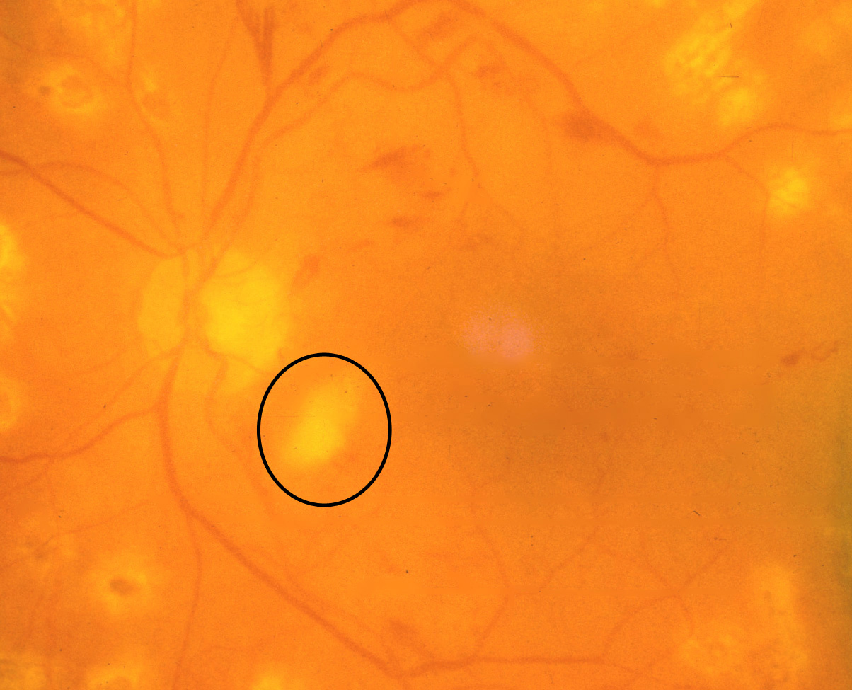
Figure 1 from Classification of Cotton Wool Spots Using Principal  Components Analysis and Support Vector Machine | Semantic Scholar
Figure 1 from Classification of Cotton Wool Spots Using Principal Components Analysis and Support Vector Machine | Semantic Scholar

Cotton wool spots. COMS Grading Scheme

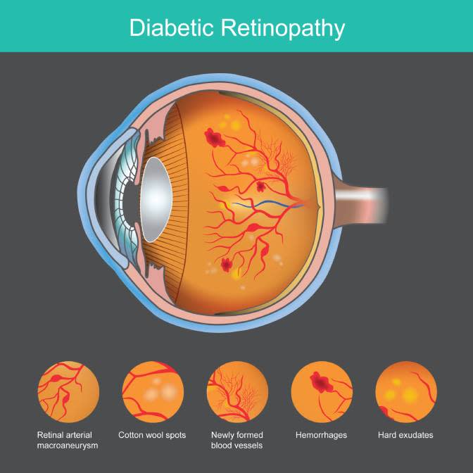
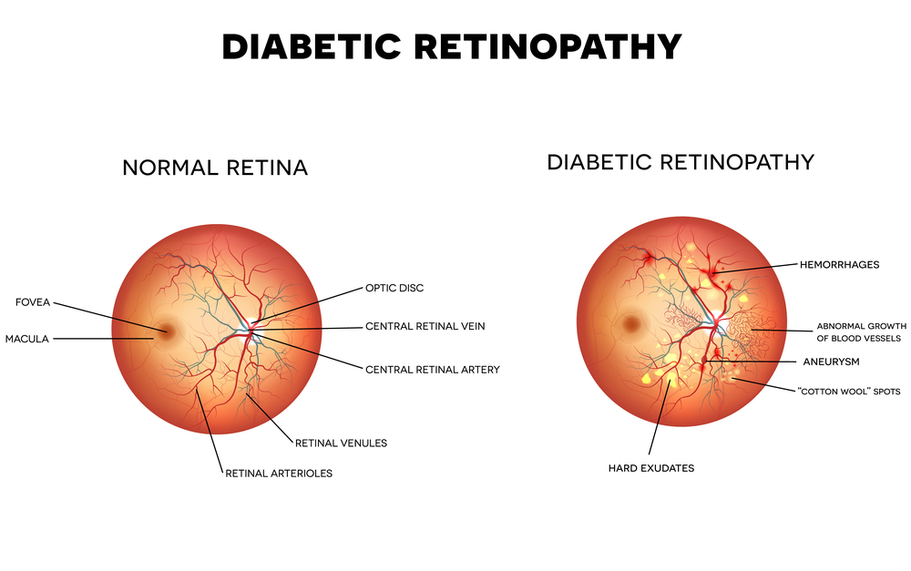
Diabetic Retinopathy Screenings | The EyeDoctors Optometrists

Diabetic Retinopathy Causes and Diagnosis | FLEI

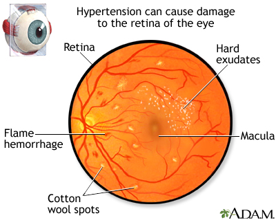
Hypertensive retinopathy: MedlinePlus Medical Encyclopedia Image

Diabetic Retinopathy Flashcards | Quizlet

Color Feature Segmentation Image for Identification of Cotton Wool Spots on  Diabetic Retinopathy Fundus
Color Feature Segmentation Image for Identification of Cotton Wool Spots on Diabetic Retinopathy Fundus

Data on fundus images for vessels segmentation, detection of hypertensive  retinopathy, diabetic retinopathy and papilledema - ScienceDirect
Data on fundus images for vessels segmentation, detection of hypertensive retinopathy, diabetic retinopathy and papilledema - ScienceDirect

Cotton-Wool Spots May Challenge Diagnosis

Symptoms of retinopathy: (a) hard exudates, (b) cotton wool spots and... |  Download Scientific Diagram
Symptoms of retinopathy: (a) hard exudates, (b) cotton wool spots and... | Download Scientific Diagram

Cotton wool spots. COMS Grading Scheme

Moran CORE | Retina & RPE Histopathology

COTTON WOOL SPOTS EXPLAINED ! - YouTube

Ophthalmology-Notes And Synopses - Layers of Retina affected in Diabetic  Retinopathy: ➖Cotton Wool Spots: Nerve fibre layer. ➖Microaneursyms: Inner  nuclear layer. ➖Dot blot hemorrhages: Inner nuclear & Outer plexiform  layer. ➖Flame-shaped hemorrhages:
Ophthalmology-Notes And Synopses - Layers of Retina affected in Diabetic Retinopathy: ➖Cotton Wool Spots: Nerve fibre layer. ➖Microaneursyms: Inner nuclear layer. ➖Dot blot hemorrhages: Inner nuclear & Outer plexiform layer. ➖Flame-shaped hemorrhages:

The Reading of Components of Diabetic Retinopathy: An Evolutionary Approach  for Filtering Normal Digital Fundus Imaging in Screening and Population  Based Studies | PLOS ONE
The Reading of Components of Diabetic Retinopathy: An Evolutionary Approach for Filtering Normal Digital Fundus Imaging in Screening and Population Based Studies | PLOS ONE

Eyes and Heart Disease | Singapore Heart Foundation

Fundoscopic Examination - ppt video online download

Fundus | Ento Key
Fundus | Ento Key

Differentiating cotton wool spot , exudates and Drusen on OCT | Eye facts,  Eye anatomy, Medical ultrasound
Differentiating cotton wool spot , exudates and Drusen on OCT | Eye facts, Eye anatomy, Medical ultrasound
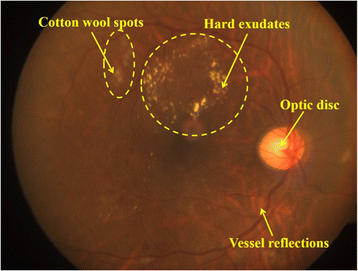
Cotton Wool Spots : Ophthalmoscopic Abnormalities : The Eyes Have It

Semi-automated quantification of hard exudates in colour fundus photographs  diagnosed with diabetic retinopathy | BMC Ophthalmology | Full Text
Semi-automated quantification of hard exudates in colour fundus photographs diagnosed with diabetic retinopathy | BMC Ophthalmology | Full Text
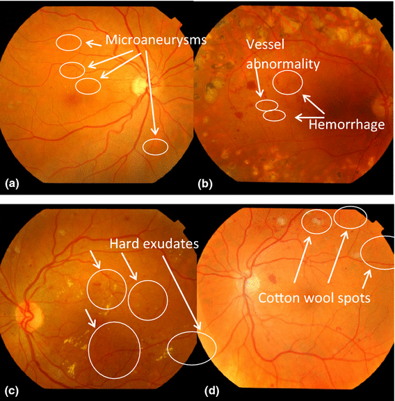
Automatic recognition of severity level for diagnosis of diabetic  retinopathy using deep visual features | SpringerLink
Automatic recognition of severity level for diagnosis of diabetic retinopathy using deep visual features | SpringerLink